 Medijoy GlobalMedijoy Global
Leveraging Boao Lecheng International Medical Pilot Zone policies, we bring world-leading diagnostic and treatment equipment to provide precise, safe, and efficient medical services.
The Da Vinci Surgical Robot is the world's most advanced minimally invasive surgery system, developed by Intuitive Surgical (USA), now in its 4th generation Xi system. With HD 3D vision, 540° rotating robotic arms, and tremor filtration technology, it surpasses the physiological limits of human hands.
3D HD Magnified View
Robotic Arm Rotation
Minimally Invasive Incision
Fewer Complications
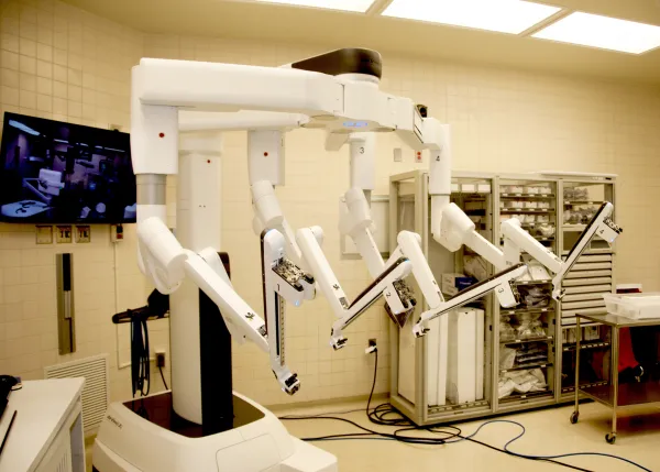
Glasses-free 3D imaging with 10-15x tissue magnification. Capillaries are clearly visible, enabling precise avoidance of critical nerves and blood vessels.
Intelligently filters natural hand tremors. Vascular accident rate reduced from 2.8% to 0.5%, with multi-fold improvement in surgical precision.
Widely used in urology, gynecology, gastrointestinal, thoracic, and hepatobiliary surgery. The preferred choice for complex procedures.
Ergonomic remote control station. Intuitive interface remembers surgeon preferences, reducing fatigue during long procedures.
4-arm bedside robotic system. Suspended arm design accesses any angle. Laser-guided positioning with automatic arm adjustment.
Glasses-free 3D HD imaging at 10-15x. Fluorescence imaging for real-time tissue perfusion. Audio feedback for critical steps.
| Metric | Open Surgery | Laparoscopic | Da Vinci Robot |
|---|---|---|---|
| Incision Size | 20-30cm | 1-2cm x 3-4 ports | 0.5-1cm x 4-5 ports |
| Blood Loss | 200-500ml+ | 50-200ml | 10-50ml |
| Vision Quality | Direct sight | 2D flat image | 3D HD 10-15x |
| Precision | Direct hand | Rigid instruments | 540deg rotation tremor-free |
| Hospital Stay | 7-14 days | 4-7 days | 2-5 days |
| Complication Rate | Higher | Moderate | 30-40pct lower |
Urology
Prostate Kidney Bladder
Gynecology
Fibroids Cysts Pelvic repair
GI Surgery
Colorectal Gastric Whipple
Thoracic
Lung Esophageal Mediastinal
HPB Surgery
Liver Gallbladder Pancreas
Cardiac
Mitral repair CABG
Head and Neck
Thyroid Throat tumors
Pediatric
All pediatric MIS
84-year-old with kidney cancer, high risk for open surgery. Robot-assisted partial nephrectomy in 2 hours, less than 50ml blood loss, walking next day, full renal preservation.
Elderly High-Risk Kidney Saved
Low rectal cancer near anal sphincter. Robot precisely removed tumor preserving function. 4cm incision, less than 10ml blood loss, discharged day 3.
Low Rectal Cancer Sphincter Saved
Whipple procedure - the Mount Everest of abdominal surgery. Robot-assisted completion with less than 50ml blood loss, thorough lymph node dissection, discharged day 10.
Complex Surgery Precision Safe
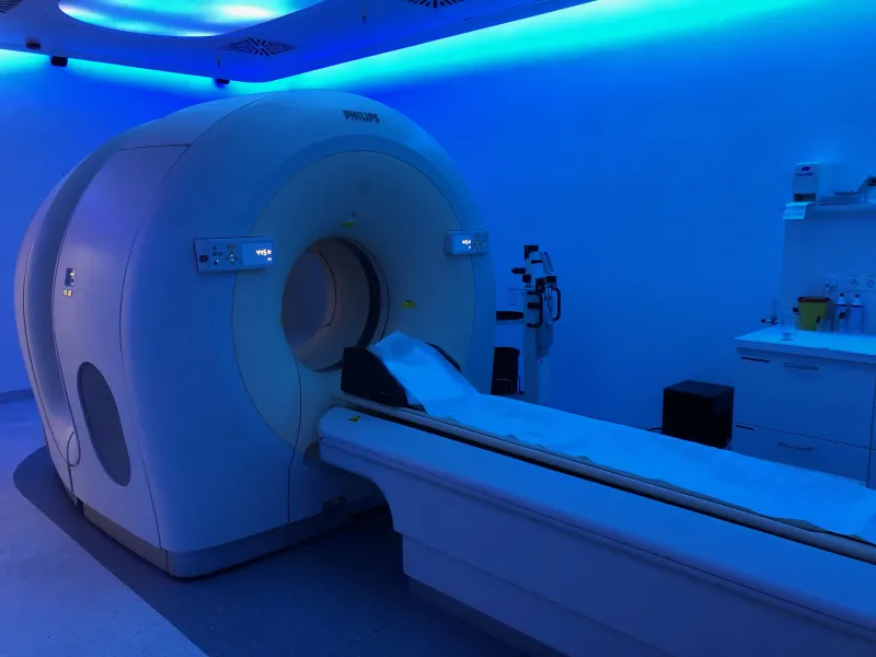
PET-CT combines Positron Emission Tomography with Computed Tomography, representing one of the world's most advanced medical imaging systems. A single scan provides both metabolic and anatomical information for early tumor detection, precise staging, and treatment assessment.
Detects Micro Lesions
Single Full-Body Scan
Cellular-Level Detection
Metabolic+Anatomical
Captures metabolic abnormalities at the cellular level, detecting lesions 3-6 months earlier than conventional CT/MRI. Full-body scan in just 20-30 minutes.
Accurately determines tumor staging and evaluates chemo/radiotherapy efficacy. One scan clarifies metastasis status, avoiding unnecessary surgeries.
Included in routine premium health checkups in Japan, Korea, and other developed countries. Ideal for those with family cancer history or long-term smoking history.
IV injection of 18F-FDG tracer (glucose analog). Cancer cells with high metabolic activity absorb large amounts, trapping the tracer. Ring detectors capture gamma photons from positron annihilation, precisely mapping metabolically active regions.
X-rays penetrate the body from multiple angles. Computer reconstruction generates high-resolution cross-sectional images showing lesion location, size, and relationship to surrounding structures, while providing attenuation correction for PET.
Family Cancer History
High-risk with family history of malignancy
Long-term Smokers
20+ years smoking, lung cancer screening
Elevated Tumor Markers
Markers elevated but routine exams negative
Diagnosed Cancer
Staging, treatment assessment, follow-up
Space-Occupying Lesions
Lesions difficult to classify benign vs malignant
Unexplained Symptoms
Prolonged fever, weight loss, pain of unknown origin
Neurological Symptoms
Epilepsy, memory decline, Parkinson-like
Coronary Disease
Post-MI myocardial viability assessment
Tracer Injection
IV injection of 18F-FDG, quick and painless
Resting Period
About 60 min for tracer body distribution
Full-Body Scan
About 20 min complete whole-body imaging
Report Reading
Multi-disciplinary expert joint report
Radiation dose about 14-20mSv per scan, below international safety limits. Tracer half-life only 109.8 min, metabolized within 24h. Drink water post-scan. Not for pregnant or breastfeeding.
Our medical advisors will recommend the most suitable examination plan and hospital based on your needs.
Inquire NowTo provide a better service, we would like to use cookies and the Meta (Facebook) pixel, with your consent, to measure site visits and optimise our advertising. You may accept or decline; declining does not affect your browsing.
See ourPrivacy Policy for details.